|
Intended Audience
A guide for researchers not familiar with latest features of the next generation 3DSuit inertial Motion Capture Systems, released in March 2012.
1. Introduction
When Inertial Labs introduced its gyroscopic
inertial motion capture system in 2006, the only
interface software that it was coupled with was
designed for use by professional animation
community who needed a less complex tool to
capture data for Animation.
Mainly due to lack of demand, there was never
any attempt by Inertial Labs to direct further
development of the technology to address needs
in the biomechanical community. This position
changed in 2010 when Inertial Labs recognized
there was an opportunity to help research
facilities and Universities as well as the animation community.
By mid-2011, Inertial Labs was able to
demonstrate its newly completed beta products to
selected researchers who were extremely
interested in helping to develop the next
generation of inertial motion capture systems.
This would include new IMU technology (Inertial Measurement Units) as well as a data acquisition pipelines covering some groundbreaking features. 3DSuit system released in March 2012 are the result of this collaboration.
This document will explain the 3DSuit systems thoroughly and help identify for the researcher in which exact tasks the Next-Gen 3DSuit systems can be employed where results will be on par with an optical system, and what quantitative tasks should not be delegated to them in the first place.
2. Gyroscopic inertial motion capture system vs. optical systems
2.1 Why compare the two systems.
Before any motion capture technology was ever commercially introduced, researchers used video cameras and high speed still cameras to capture images in calibrated angles of view and then attempt to line body segments to calculate joint angles. This was (crude and tedious but effective.
This process of arriving at joint angles from digitizing body segments is ingrained in the biomechanics community’s understanding of how angle data is digitized. From this point of view, any system contending to be a biomechanical tool needs to be compared against the general optical system pipeline of collecting data.
When evaluated against optical systems, Gyroscopic system manufacturers have consistently failed to make a positive impression, even though they employ one of the most accurate sensors ever developed.
They have not helped their case by offering their systems as a 6 Degrees of Freedom (6DoF) alternative to optical systems since in such a configuration, with large tolerance positional data (either measured or calculated), the inertial systems tend to corrupt the accuracy of their rotational data.
The accurate rotational data when left alone can be a valuable tool for a wide selection of research projects.
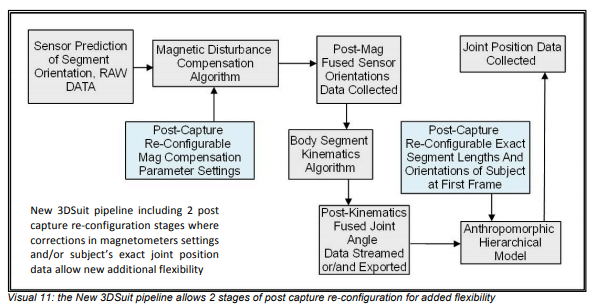
The new 3DSuit systems don’t compete with optical pipelines, but try to complement them. In situations where it is the only candidate, it increases flexibility through its optional user configured (and reconfigurable in post) data file of exact subject segment lengths & orientations. This file also holds reconfigurable magnetic compensation settings for the Raw data – an industry first.
Seasoned optical system users have the best tool for capturing data indoors (lab conditions) and have experienced quality data. Those same users can now get similar data on a real bike, in a real car, on a sandy beach, in a kayak, at a house, or running on the grass and this is why we think they would be very interested in adding the 3DSuit system to their research tools.
2.2 Main system advantages and drawbacks
Some general attributes of inertial and optical motion capture systems are tabled below. Note that in the following table only high-end optical systems, as well as the Next-Gen 3DSuit systems are considered.
 2.3 Shared challenges of the 2 systems 2.3 Shared challenges of the 2 systems
Regardless of the type of system used there remain a few challenges which the user needs to learn to mitigate if they want to maximise the accuracy of collected data.
1. Clothing artefact: clothes stretch and change position. The value of accurate sensors is negated if they move with clothes rather than the body
2. Tissue artefact: there are no solid spaces on the thighs and upper arms to place any markers or sensors. Markers/sensors placed on these segments will move when their distal segments move, even if the segment itself has not moved.
3. Skin artefact: sensors need to move with the body to provide accurate data. Sensors moving on loose skin will introduce data artefacts. The size and weight of sensor will reduce this effect, so this is more of a challenge for the inertial system, requiring the IMUs be the lightest and smallest possible size and weight.
4. Kinetic artefact: the momentum of a fist will move sensors/markers beyond their intended travel. This again, is more of a challenge for inertial systems (even though the IMUs are strapped on segments) so size and weight of IMUs are a significant factor in choosing a motion capture system.
2.4 Differences in the approach to data collection (Lines v dots)

Both systems are primarily used to collect joint angle data however they go about it in a very different way. Optical systems measure limb position displacement in reference to a user-designated origin, and thereafter calculate the angle of the said limb against the same origin (or another limb).
Inertial systems measure limb orientation displacement directly, bypassing the need for segment position data. An inertial system, however, needs an anthropomorphic model at the outset on which to display the motions.
The segment lengths of the said model do not alter the accuracy of angles between them (joint positions, however, would be arbitrary). With accurate segment lengths and orientations at the start, either measured manually or photographically, a layer of accuracy would apply to joint positions in addition to already accurate joint angles.
Both systems start off with global coordinates, 3DSuit uses Earth’s magnetic field and gravity as reference and cameras designate an origin somewhere in the capture area (hence 3DSuit can have an extremely large capture area).

2.5 Optical v inertial anthropomorphic model
Another difference between the 2 systems is how they display their data. Optical systems, create a representation of the subject in the first few frames of capture which will be an exact graphical display of the collected data, with accurate segment positions. Inertial systems on the other hand need a pre-designated anthropomorphic model in place to assign the collected orientation data to the corresponding segments.
The model used by 3DSuit systems is a hierarchical kinematic chain of segments starting at the Pelvis segment (with the root of the hierarchy at its centre and positioned directly above Global coordinate origin). All other joints are linked to the root through 5 clusters of 3 to 5 IMUs (segments) each.
The main characteristic of a hierarchical model is that all joints are parented by another joint (except the root which is parented by Global origin). Any angular displacement attributed to a parenting joint will affect the position of its children.


2.6 Optically Configured 3DSuit Model for Accurate Local Positional Data
Some inertial systems use statistical anthropomorphic models, which are partially scalable and are matched to the subject’s stance at the start of data stream. This system can’t ensure accurate joint positions, making all joint linear acceleration and displacement data questionable.
3DSuit uses front and side photos of the subject at natural (and repeatable) rest posture while inside a cube with known dimensions and uses a drag-n-drop method of determining the exact joint positions (and segment orientation) of the subject.
This file (Actor file) containing the subject’s size and posture can be modified after data collection and re-fused into the collected sensor data in post. This method has the following benefits:
1. Provides accurate local positional data (relative to root, not the global origin)
2. Solid footsteps
3. Start data stream at any position e.g. Sitting position (ideal for disabled, elderly, patients)
4. Make animal or human section models easily
5. Separate and independent from collected data (save session time, use default model, take photos only, create and input Actor file later)
6. Correct mistakes, re-tune Actor file in post
  
2.7 Grouping of segments and joints of the 3DSuit model in levels of angular data accuracy
One of the best ways to take on any job is to understand and take stock of available tools.
One of the changes in the approach to data collection adopted by the new 3DSuit systems is to group segments and joints into 3 sets based on the level of angular data accuracy obtainable by each. The 3 groups are separated and named by Inertial Labs as follows:
Alpha segments and joints with high level of Raw and Fused data accuracy expected under most every condition and range of movement
Beta segments and joints can only obtain the same level of expected Raw and Fused data accuracy as Alpha ones, if they are either employed for the capture of limited range of movements, or if the sensors are placed on segments using rigid plates fastened at both ends of the segment in order to be unaffected by the segment’s tissue artefact
Complex segments and joints are unable to achieve high levels of Raw or Fused data accuracy expectancy since they are generally encompass multiple numbers of bones working together in various axes but are measured using a single sensor which will measure more of the displacement incurred by the bone nearest to it.
 The level of accuracy is determined by various factors: The level of accuracy is determined by various factors:
- The amount of noise generated by the placement of the IMU (due to clothing stretch, skin instability, and IMU kinetic artefact produced with sudden changes)
- Unavailability of proper amount of space on the segment to place IMUs (tissue artefact from the motion of distal segments
- Complex skeletal segments represented by a single sensor (Spine, clavicle)
NOTE: The accuracy attributed to Alpha joints is not impaired even though they usually have other types of segments connecting them to the origin. This is due to the fact that both segments that make up the joint use the same subtraction to the origin and are left only with their own orientation.
2.8 Can Inertial systems provide 6DoF data?
In their push to provide a complete data set (6DoF), inertial system pipelines employ various methods of adding positional information to their collected orientation data but in doing so they trade off their accurate 3DoF data with a not so accurate 6DoF data set.
To get around this unnecessary dilution of perfectly good data, the new 3DSuit systems offer the following new flexibilities:
- Absolute orientation of segments (Raw Data) before they are fused with magnetic disturbance compensation (MDC) algorithm
- Post MDC algorithm fused data before it is fused with kinematics algorithm
- User re-configurable MDC algorithm parameter settings
- User re-configurable joint position data (in post) for accurate local positional data and more accurate global positional 6DoF dataset
- Software to allow establishment of subject’s joint positions at rest (using photos)
- Simultaneous stream and collection of post magnetic compensation fused Quaternion data and post kinematics fused Euler angles (both re-configurable in post)
- Grouping of segments into 3 levels based on level of angular data accuracy obtainable

The above features help the researcher to determine if the 3DSuit can be an accurate tool to collect the particular type of data they need. In many instances what they need is provided by the 3DSuit system without going 6DoF and positional information is never used throughout the pipeline.
3. New 3DSuit technologies and pipelines suited to Biomechanical applications
Based on market research and a focussed R&D effort, the following series of hardware changes are available in the Next-Gen 3DSuit systems:
3.1 Miniaturization of IMUs and reduction of other hardware footprint
- Small (Heavy Duty 48 x 16 x 9 mm or 7cm3) IMUs drastically reduce footprint effects of kinetic forces In fact action, helping Secondary Beta segments capture data quality on par with Primary Beta segments
 
- Separation of Main Processing Unit from its battery and wireless units allows users to use the lighter size batteries when lengthy sessions are not intended (largest size battery can last 13 hours and smallest 2 hours). Sometimes wired systems are more apt for some situation.
- Patent pending Splitter Boxes can spread the connections around the suit
- TTL Signal hardware Sync box is external and smaller than a matchbox
- Smaller components means new smaller carrying cases
3.2 Configurable kinematics model in post
As outlined in previous section positional data from inertial systems are always somewhat inaccurate but 3DSuit provides a stand-alone software package that uses 2 photos of the subject and an easy drag-n-drop system to configure the size and orientation of the of the subject’s segments for more accurate positional data
3.3 Synchronised fused and raw real-time data collection (in addition to TTL synch)
3.3.1. This new feature of the 3DSuit systems will reduce the impact of magnetic disturbance on IMUs. 3DSuit systems as standard will capture 2 sets of synchronised data, one, data fused with magnetic compensation AND kinematics algorithm (to see a representation of the data), and another, data fused only with magnetic compensation algorithm (Raw Data). The new feature is that the Raw Data will have magnetic state of each IMU in every frame, which can be matched to anomalous segment behaviour on the anthropomorphic model in the post kinematics data.
3.3.2. Once erroneous magnetic compensation settings are recognised in the Raw Data, the settings can be altered and the raw data re-computed for another attempt in finding the perfect compensation settings parameters
3.3.3. Not only this process can be repeated until satisfied with the results, but can be split for every frame of set of frames, or IMUs in a frame or set of IMUs: as detailed as the magnetic disturbance requires it to be.
3.4 Data acquisition window
Feature to manage naming and organizing files of collected data with ease
3.5 New biomechanical Graphical User Interface
Includes multi windows to see model and graphs together in real-time
3.6 A TTL hardware synch system.
This system can be configured to connect to any DAQ card in the market
3.7 Cloth Suit Technology
We have 19 years of experience in how to design clothing to reduce artefacts in collected data and we have introduced a new 4-way stretch suit in addition to the Basic and the Strap suits. It is designed for special medical or biomechanical cases when disabled subjects need assistance to wear the system.
3.8 No 3DSuit system needs longer than 3 seconds to start
This also includes a fresh set of calibration for all the IMUs at every start
4. DATA EXPORT FORMATS
4.1 Raw data
The data output in this format is in 2 groups. The
collected sensor data at the time of capture (real
raw data) and the post-magnetic disturbance
compensation algorithm (first stage fused data).
What is revolutionary about this data file is that
the ‘Post-Mag’ algorithm section can be reconfigured
by changing the setting in the ini file
and re-computing it. The first part of the data
including the raw magnetometer, gyroscope and
accelerometer data will stay the same but a new
magnetic algorithm parameter setting will change
the quaternion and Euler angles within the file.
NOTE: this Raw Data file can and usually is, recorded in synch with an accompanying file recording post magnetic compensation and post kinematics algorithm Fused Data output
  4.2 Second Stage Fused data (bvh) 4.2 Second Stage Fused data (bvh)
The Fused Data in this file has not only gone through the first stage fusion (magnetic compensation fusion) but also the second stage; the kinematics algorithm fusion.
This file, when the data collected is mostly raw, can be used as a reference to see how the various segments are holding up to magnetic interference in the capture space. If this data shows magnetic disturbance, the researcher can go back to the Raw data and infuse the ini file with new setting for the magnetic algorithm to try to compensate the disturbance fully.
Each time, the result of the re-configuration of the settings can be viewed in the file (after it has been re-computed in the Raw data file).
The bvh data (biovision hierarchy model) provides 2 main sections in the output file.
The first is the exact offsets for the anthropomorphic model segment lengths (in inches) and orientations by supplying the joint positions.
The second section is 66 columns of data. The first 6 are the positional and angular displacement of the centre of pelvis (Root) in reference to the global origin, and the next 60, are the angular displacements of 20 joints, all traced back to the root either directly of through other linked joints. The order of axes in this file for every joint is Z X Y (but XYZ for the first 3 which are positional data).
The joint coordinate system on the bvh is Y up (the 3DSuit model JCS turned about the X, 90 degrees plus which makes: 3DSuit model +X = bvh +X, +Y = -Z, +Z = +Y.


4.3 Cross reference table
The following table shows the matching of the various joint coordinate systems used as well as the direction of angular displacement in the bvh data matching to biomechanical terminology for the action, as well as the column number of the data in bvh in which each joint action is recorded can be found in the following comprehensive table.

4.4 Fused positional data (.txt)
There is a text format file output of the joint positions which is self simply lists the Joint names and their XYZ positions below the name respectively. The Joint coordinate system used is the same as the 3DSuit kinematics model (shown in visuals 6 and 17)

4.5 Data Converters
- XML format
- C3D format
|